Instruction Manual
46 Pages
Preview
Page 1
ENGLISH
INSTRUCTION MANUAL
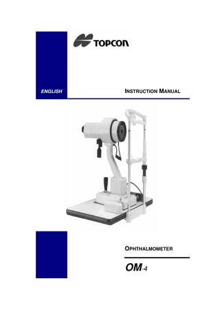
OPHTHALMOMETER
OM-4
INTRODUCTION
Thank you very much for purchasing the TOPCON Non-Mydriatic Ophthalmometer OM-4. This Instruction Manual gives a description of the TOPCON OM-4. This includes its main features and basic operation, troubleshooting and the checking, maintenance and cleaning of this instrument. To get the best use from this instrument, please carefully read these instructions and place this manual in a convenient location for future reference.
Ophthalmometer OM-4
Precautions
● This Ophthalmometer is a piece of precision equipment, which needs
● ●
● ●
●
●
to be used and kept in places under normal life conditions regarding temperature and humidity. Do not expose the instrument to direct sunshine. To ensure best use, install the instrument on a level floor free from any vibration. Always check that the power cord is plugged in correctly before use. WARNING: For safety always make sure the instrument is correctly grounded. Use care that no fingerprints or foreign matter remains on the objective lens. Topcon is not responsible for any modification caused by disassembling or adjustments made by unauthorized dealer or persons. If any trouble occurs with your instrument or its accessories, first refer to the troubleshooting guide in this manual and carry out the checks listed there. If nothing is found during your check, then contact your authorized dealer or TOPCON to service it. Always turn the power source off and place the dustcover on the instrument when it is not in operation.
Note The TOPCON Ophthalmometer OM-4 has the CE mark, when used in combination with its original power supply, chin rest and table-top. In case one (or more) of the above two elements is (are) not used, please note the following must be done to remain CE compliant; ● Another power supply needs to have a CE mark, and the following
specification. Voltage at the lamp in putting on the 6V3W halogen lamp; Max 7.5V ● The total installation needs to be earthed.
iv
Introduction
Displays for Safe Use In order to encourage the safe use of products and to prevent any danger for the operator and others, or damage to properties, important warning signs are placed on the products and mentioned in the instruction manuals. We suggest that everyone understands the meaning of the following displays and icons, before reading the “Safety Precautions” and text in this manual.
Display
Meaning
Ignoring or disregarding this display may lead to personal injury or severe damage to the instrument or facilities. CAUTION ● Injury potential includes hurt, burn, electric shock, etc. ● Damage to facilities refers to extensive damage to buildings,
equipment and furniture.
Icons
Meaning This indicates Hazard Alert (Warning). Specific content is expressed with words or an image, either inserted in the icon itself or located close to the icon.
INSTRUCTION MANUAL
v
Ophthalmometer OM-4
Safety Precautions
CAUTION Icons
Prevention item
To prevent bumping the machine against the patient's face, pay special attention when operating the equipment.
To prevent your fingers getting caught, pay special attention when operating the equipment.
To prevent causing pain to the patients or injury to their eyes, never raise the brightness more than necessary.
To prevent an electrical shock, turn off the power switch and disconnect the power cord before replacing lamps.
To prevent an electrical shock, turn off the power switch and disconnect power cord before replacing fuses.
To prevent heat burns, pay special attention when replacing lamps, since the lamp unit and the lamp housing are red hot.
To prevent dropping possible accessories during use or during relocation, fasten the fixing screws firmly.
vi
Introduction
Operation and Maintenance Usage The Ophthalmometer is a piece of medical equipment and the operator must be a properly trained and experienced doctor.
Escape clause
● TOPCON shall not take any responsibility for damage due to fire,
earthquakes, actions by third persons and other accidents, or the negligence and misuse by the user and use under unusual conditions. ● TOPCON shall not take any responsibility for damage derived from the inability to use this equipment, such as a loss of business profit and suspension of business. ● TOPCON shall not take any responsibility for damage caused by any oyher operation than that described in this Instruction Manual. ● Diagnoses shall be made on the responsibility of the doctors concerned and TOPCON shall not take any responsibility for the results of such diagnoses.
INSTRUCTION MANUAL
vii
Ophthalmometer OM-4
Warning indications and positions To insure safety, warning labels are provided. Use the equipment correctly according to these warning instructions. If any of the following warning labels are missing, please contact us at the address stated on the back cover of this manual. CAUTION • To prevent an electrical shock, turn off the power switch and disconnect the power cord before replacing lamps. • To prevent heat burns, pay special attention when replacing lamps, since the lamp unit and the lamp housing are red hot.
CAUTION • To prevent bumping the machine against the patient's face, pay special attention when operating the equipment. • To prevent your fingers getting caught, pay special attention when operating the equipment.
CAUTION • To prevent an electrical shock, turn off the power switch and disconnect power cord before replacing fuses.
viii
CONTENTS
INTRODUCTION Precautions
iv
Note... iv
Displays for Safe Use
v
Safety Precautions
vi
Operation and Maintenance
vii
Usage...
1
vii
Escape clause
vii
Warning indications and positions
viii
COMPONENTS 1.1
Component names
12
1.2
Assembly Components
14
2
ASSEMBLY 2.1
Assembly Procedure
17
2.1.1
Installing the Table... 17
2.1.2
Installing the ophthalmic stand... 18
2.1.3
Attachment of the chin rest and headrest section... 18
2.1.4
Setting up the Base... 19
2.1.5
Fixing the Chin Rest Pads... 19
Ophthalmometer OM-4
2.1.6
Connecting the Electrical Cords... 20
2.1.7
Check the light... 21
3
OPERATION PROCEDURE 3.1
Preparations
23
3.2
Practice measurements with the test ball
25
3.2.1
Attachment of the column... 25
3.2.2
Position the instrument correctly before the test ball... 25
3.2.3
Measurement... 27
3.3
Positioning the patient... 28
3.3.2
Positioning the instrument in front of the patient... 29
3.3.3
Measurements... 30
x
Measurement of contact lenses
32
3.4.1
Attachment of the column... 32
3.4.2
Correct position of the instrument to the contact lens... 32
3.4.3
Measurement... 33
4
6
28
3.3.1
3.4
5
Measurement of the radius of the cornea curvature
MAINTENANCE 4.1
Changing the lamp
35
4.2
Exchanging the fuse
36
4.3
Cleaning the instrument
36
4.4
General care
37
4.5
Before requesting service
37
FEATURES Internal reading scales
39
Wide range of measurements
39
Highly precise measurements
40
Speedy and accurate focussing adjustments
40
Speedy one-position measurement
40
Superior handling ease
40
Inspection of an unusual cornea in irregular astigmatism
41
SPECIFICATIONS
1
COMPONENTS
15
14
16
13 17
12 10
11
9 8
1
2
7
6 5
3 4
Ophthalmometer OM-4
1.1 Component names
12
(1)
Connector cord
(2)
Table
(3)
Powercord
(4)
Power switch
(5)
Illumination control switch
(6)
Power supply unit
(7)
Gliding plate
(8)
Omni-directional joystick
(9)
Axis rotating handle
(10)
Auxilary knob
(11)
Horizontal knob
(12)
Vertical knob
(13)
Eyepiece
(14)
Vertical index
(15)
Protractor scale
(16)
Levelling pin
(17)
Horizontal index
(18)
Rail
(19)
Chin rest adjustment knob
(20)
Chin rest
(21)
Occluder
(22)
Headrest
(23)
Mire plate
(24)
Height mark
Components
23
22
21
24
20 19
18
Contact lens holder Test ball (R 7.50)
Column
INSTRUCTION MANUAL
13
Ophthalmometer OM-4
1.2 Assembly Components The following items are packaged in a box
Figure 1
Figure 2
14
Accessories
Quantity
Instruction manual
1 each
Compensation table
1 each
Dust cover
1 each
Table top
1 each
Accessory box
1 set
Rail covers
1 pair
Power cord
1 each
Chin rest and head rest
1 each
Screwdriver
1 each
Measuring head and base
1 each
Components
The contents of the accessory box are as follows
Accessories
Quantity
Spanner (not supplied with the table for ophthalmic stand)
1 each
Silicone cloth
1 each
Lamps (spares)
9 each
Contact lens holder
1 each
Test ball (R 7.50)
1 each
Fuse
2 each
Chin rest pads
1 pack
Column
1 each
Cleaning brush
1 each
Pad fixing pin
2 each
Figure 3
INSTRUCTION MANUAL
15
Ophthalmometer OM-4
16
2
ASSEMBLY
These instructions are for assembling the OM-4 Ophthalmometer after all the components have been carefully removed from the shipping carton.
2.1 Assembly Procedure Please check the primary voltage set for the instrument. (see Figure 9)
2.1.1
Installing the Table Unscrew the four attachment bolts underneath the instrument table. These bolts have only been screwed into their holes lightly and can be very easily unscrewed. Use them to fix the table on to the elevating mechanism of the adjustable instrument table, as illustrated in Figure 4.
Ophthalmometer OM-4
Figure 4
2.1.2
Installing the ophthalmic stand Simply insert the pole protruding from the surface of the table into the socket on the lower instrument arm and tighten the fixing screw, as shown in Figure 5.
Figure 5
2.1.3
Attachment of the chin rest and headrest section Unscrew the four attachment screws, which have been lightly screwed into the attachment mount of the chin rest and headrest section. Then fix the section to the mount with these four screws, as shown in Figure 6.
18
Assembly
Figure 6
2.1.4
Setting up the Base 1 Simply place the base section (with the measuring head) on top of the table, with the outrigger rollers aligned on top of the rail teeth (18). 2 Then place the rail covers over the rails, by inserting the flange of the cover into the slight opening between the rail and table surface, as in Figure 7.
Figure 7
2.1.5
Fixing the Chin Rest Pads 1 Pull out the pad fixing pins of the chin rest (20), by pushing them up. 2 Next, place a suitable quantity of pads on the chin rest and fix them in place with the two pad fixing pins, as in Figure 8.
INSTRUCTION MANUAL
19
Ophthalmometer OM-4
Figure 8
Tear off one tissue pad after each patient, thereby uncovering a new one for the next.
2.1.6
Connecting the Electrical Cords 1 Switch off the power switch (5). 2 Then, connect the power cord (3) and connector cord (1) to the room receptacle and connector respectively, as in Figure 9 and Figure 10.
Figure 9
Voltage selector
Figure 10
20
Assembly
2.1.7
Check the light Switch on the power switch and check whether the pilot lamp, mire lamp and illumination lamp for the scale light up.
INSTRUCTION MANUAL
21