Instruction Manual
80 Pages
Preview
Page 1
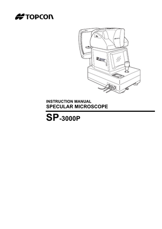
INSTRUCTION MANUAL
SPECULAR MICROSCOPE
SP-3000P
INTRODUCTION Thank you for purchasing the TOPCON SP-3000P Specular Microscope.
This instrument is used to photograph and evaluate the corneal endothelium. This instrument has the following features: • Photography of the corneal endothelium and the measurement of the corneal thickness can be performed at the same time. • The auto-alignment function ensures quick and easy centering and measurement. • Simplified cell analysis enables the user to easily understand the rough value of the cell density.
This manual outlines the SP-3000P Specular Microscope, its operating procedures, trouble shooting, maintenance and cleaning. Before using this instrument, carefully read the "DISPLAY FOR SAFE USE" and the "SAFETY CAUTIONS" to familiarize yourself with the features of the SP-3000P Specular Microscope and to ensure that you operate it efficiently and safely. Always keep this instruction manual at hand.
[Warning] Care should be taken not to hit the patient's eye or nose with the instrument during operation. (The patient may be injured.)
This symbol is applicable for EU member countries only. To avoid potential negative consequences for the environment and possibly human health, this instrument should be disposed of (i) for EU member countries - in accordance with WEEE (Directive on Waste Electrical and Electronic Equipment), or (ii) for all other countries, in accordance with local disposal and recycling laws.
1
CAUTIONS FOR USE Basic caution When moving the chinrest up and down, be careful not to catch the patient's hand. The patient may be injured. To avoid electric shock, do not open the instrument. Request service from an authorized Topcon distributor. Electric shock may cause burns or a possible fire. Turn the power switch OFF and unplug the power cord before replacing the fuses. Replace only with fuses of the correct rating.
Disposal Dispose of the instrument according to local disposal and recycling laws.
ENVIRONMENTAL CONDITIONS FOR USE Temperature : 10°C ~ 40°C Humidity : 30% ~ 75% (without dew condensation) Air pressure : 700hPa ~ 1060hPa
STORAGE, USAGE PERIOD AND OTHERS 1. Environmental conditions for installation (without package) Temperature : 10°C ~ 40°C Humidity : 30% ~ 75% (without dew condensation) Air pressure : 700hPa ~ 1060hPa 2. When storing the instrument, ensure that the following conditions are met: (1) The instrument should not be splashed with water. (2) Store the instrument where air pressure, temperature, humidity, ventilation, sunlight, dust, salty/ sulfurous air, etc. do not give any negative side effect. (3) Do not store or transport the instrument on a slope or uneven surface or in an area where it is subject to vibrations or instability. (4) Do not store the instrument where chemicals are stored or gas is generated. 3. Usage period 8 years from delivery providing regular maintenance is performed (according to the self-certification [Topcon data])
ENVIRONMENTAL CONDITIONS FOR PACKAGING IN TRANSPORTATION / STORAGE Temperature : -20°C ~ 50°C Humidity : 10% ~ 95%
CHECKPOINTS FOR MAINTENANCE 1. Periodically inspect the instrument and its parts. 2. Before using the instrument after a long period of inactivity, make sure that it operates safely and normally. 3. Be careful not to stain the photography window with fingerprints or dirt. This may affect picture quality. 4. If the photography window is stained, clean it according to the "CLEANING THE PHOTOGRAPHY WINDOW" instructions on P. 74 in this manual.
2
DISPLAY FOR SAFE USE To encourage safe and proper use and to prevent danger to the operator and others or potential damage to property, important cautionary messages are placed on the instrument body and inserted in the instruction manual. We suggest that everyone using the instrument understands the meaning of the following displays, icons and text before reading the "SAFETY CAUTIONS" and observe all listed instructions.
DISPLAYS Display
Meaning
WARNING
Incorrect handling by ignoring this display may lead to a risk of
CAUTION
Incorrect handling by ignoring this display may lead to per-
death or serious injury.
sonal injury or physical damage.
• Injury refers to cuts, bruises, burns, electric shock, etc. which do not require hospitalization or extended medical treatment. • Physical damage refers to extensive damage to the building, nearby equipment and/or surrounding furniture.
ICONS Icon
Meaning Prohibition: Specific content is expressed with words or a picture near the icon. Mandatory Action: Specific content is expressed with words or a picture near the icon. Caution: Specific content is expressed with words or a picture near the icon.
3
SAFETY CAUTIONS WARNING Icon
Prevention item
Page
To avoid fire and electric shock in case of leakage, be sure to use a power supply equipped with a 3-plug AC receptacle for proper grounding.
16
Electric shock may cause burns or a possible fire. Turn the power switch OFF and unplug the power cord before replacing the fuses. Replace only with fuses of the correct rating.
71
CAUTION Icon
4
Prevention Item
Page
To avoid injury and/or damage to the instrument, hold the instrument in the specified position. Be careful not to shake or move the instrument suddenly.
15
When carrying the instrument, be careful not to drop or tip it over. Be careful to prevent your hand from being pinched by the instrument. If not, you may be injured.
15
To avoid electric shock, do not handle the plugs with wet fingers.
16
To avoid injury, do not let the patient put his/her hand under the chinrest.
34
Don't put your hand under the measuring head. * Instruct the patient properly. When the measuring head is lowered, it may injure you by pinching your hand.
37
To avoid injury during operation, be careful not to hit the patient's eye or nose with the instrument.
36, 41 45
Before handling the chinrest manually in the case of a malfunction, remove the power cord from the instrument. If not, your finger may be pinched and injured by a wrong operation.
58
To avoid damage to the instrument or electric shock, turn the power switch OFF and unplug the power cord before replacing the xenon lamp. Do not use any other types of xenon lamps except those specified in this manual.
69
Icon
Prevention Item
Page
To avoid damage to the instrument or electric shock, turn the power switch OFF and unplug the power cord before maintenance.
73
To avoid electric shock, do not open the instrument. Request service from an authorized Topcon distributor.
-----
This instrument has been tested (with 120V/230V) and found to comply with IEC60601-1-2: 2001. This instrument radiates radio frequency energy within standard and may affect other devices in the vicinity. If you have discovered that turning on/off the instrument affects other devices, we recommend you change its position, keep a proper distance from other devices, or plug it into a different outlet. Please consult your authorized dealer if you have any additional questions.
-----
5
USAGE AND MAINTENANCE USAGE Usage • The SP-3000P Specular Microscope is an electronic instrument for medical use. Use this instrument under a doctor's guidance.
USER MAINTENANCE To ensure the safety and performance of the instrument, all maintenance work, unless specified in this manual, shall be conducted by trained service personnel. The following maintenance tasks may be done by the user. For details, see the appropriate part of this manual.
Replacing the fuse The fuse in the instrument body may be replaced by the user. For details, refer to P.71.
Replacing the xenon lamp The xenon lamp for photography may be replaced by the user. For details, refer to P.69.
Cleaning the photography window The lens surface and glass surface of the photography window may be cleaned by the user. For details, refer to P.74.
ESCAPE CLAUSE • TOPCON shall not take any responsibility for damage due to fire, earthquakes, actions by third persons and other accidents, or damage due to negligence and misuse by the user and any use under unusual conditions. • TOPCON shall not take any responsibility for damage derived from inability to properly use this instrument, such as loss of business profit and suspension of business. • TOPCON shall not take any responsibility for damage caused from using this instrument in a manner other than that described in this instruction manual. • Diagnoses made shall be the responsibility of the pertaining doctors and TOPCON shall not take any responsibility for the results of such diagnoses.
6
WARNING DISPLAYS AND POSITIONS To ensure safety, the instrument provides warning displays. Use the instrument correctly by observing the display instructions. If any of the following display labels are missing, contact your dealer or TOPCON at the address on the back cover.
CAUTION To avoid injury, be careful not to pinch the patient's hand when operating the chinrest up/down switches.
CAUTION To avoid injury during operation, be careful not to hit the patient's eye or nose with the instrument.
NOTICE When the room temperature is 10°C or less, turn on the power 30 minutes before using the instrument to maintain the detection accuracy.
CAUTION To avoid electric shock, do not open the instrument. Request service from an authorized Topcon distributor.
WARNING Electric shock may cause burns or a possible fire. Turn the power switch OFF and unplug the power cord before replacing the fuses. Replace only with fuses of the correct rating.
7
CONTENTS CAUTIONS FOR USE ... 2 ENVIRONMENTAL CONDITIONS FOR USE ... 2 STORAGE, USAGE PERIOD AND OTHERS ... 2 ENVIRONMENTAL CONDITIONS FOR PACKAGING IN TRANSPORTATION / STORAGE ... 2 CHECKPOINTS FOR MAINTENANCE ... 2 DISPLAY FOR SAFE USE ... 3 SAFETY CAUTIONS ... 4 USAGE AND MAINTENANCE ... 6 USAGE ... 6 USER MAINTENANCE ... 6 ESCAPE CLAUSE ... 6 WARNING DISPLAYS AND POSITIONS ... 7
NOMENCLATURE ... 10 MAIN COMPONENT NAMES ... 10 COMPOSITION OF PARTS WHICH CONTACT THE HUMAN BODY ... 10 CONTROL PANEL ... 11 MONITOR SCREEN ... 11 STANDARD ACCESSORIES ... 14
PREPARATONS BEFORE USE ... 15 INSTALLING THE INSTRUMENT ... 15 CONNECTING THE POWER CORD ... 16 CONNECTING THE MOUSE ... 16 CONNECTING THE EXTERNAL INPUT/OUTPUT TERMINAL ... 17 DEFAULT SETTING ... 19 A VARIETY OF SETTINGS ... 27 RESET FROM POWER SAVE STATUS ... 33
BASIC OPERATION ... 34 PREPARATION BEFORE PHOTOGRAPHY ... 34 PHOTOGRAPHY IN AUTO MODE ... 35 PHOTOGRAPHY IN SEMI-AUTO MODE ... 40 PHOTOGRAPHY IN MANUAL MODE ... 44 DISPLAY AND DELETION OF IMAGES ... 48
OPERATION BY OBJECTS ... 50 SIMPLIFIED CELL ANALYSIS ... 50 OUTPUT TO THE IMAGEnet system ... 53 OUTPUT TO THE IMAGE PROCESSING UNIT ... 53 INPUT/OUTPUT THROUGH RS-232C ... 54
TROUBLE SHOOTING ... 57 TROUBLE SHOOTING GUIDE ... 57 OPERATING THE CHINREST MANUALLY IN THE CASE OF A MALFUNCTION ... 58
SPECIFICATIONS & PERFORMANCE ... 59 SPECIFICATIONS ... 59 ELECTROMAGNETIC COMPATIBILITY ... 60 Information about the optical radiation hazard for the user ... 64 ELECTRIC RATING ... 65 SYSTEM CLASSIFICATION ... 65 DIMENSIONS AND WEIGHT ... 65 PURPOSE OF USE ... 65 OPERATION PRINCIPLE ... 66 RELATED PRODUCT ... 66 RS-232C COMMUNICATION SPECIFICATIONS ... 67
8
MAINTENANCE AND INSPECTION ... 69 MAINTAINING ACCURACY ... 69 MAINTENANCE AND INSPECTION ... 72
MAINTENANCE ... 73 CLEANING THE DUST COVER, CONTROL PANEL, MONITOR SCREEN, ETC. ... 73 CLEANING THE PARTS WHICH COME INTO CONTACT WITH THE PATIENT ... 73 CLEANING THE PHOTOGRAPHY WINDOW ... 74
REFERENCE MATERIAL ... 75 OPTIONAL ACCESSORIES ... 75
TERMINOLOGY ... 76 DESCRIPTION OF TERMS ... 76
9
NOMENCLATURE MAIN COMPONENT NAMES Lamp house cover
Photography head unit Color video monitor
Photography switch
Base unit
Joystick
Brightness volume Control panel
Power lamp RS-232C input terminal
Power supply unit VIDEO OUT output terminal USB output terminal
Locking knob (for movement)
Forehead rest Photography window Vertical positioning mark
Chinrest tissue pin
Power switch
Chinrest
Chinrest unit Fuse holder
Power cord
COMPOSITION OF PARTS WHICH CONTACT THE HUMAN BODY Forehead rest : Silicone rubber Chinrest
10
NOMENCLATURE
: Acrylonitrile butadiene styrene resin
CONTROL PANEL
Clear switch
Delete switch
Menu switch Image selector switch
Image transfer switch Chinrest down switch
Chinrest up switch
Power LED
Delete switch
: Deletes the image being displayed.
Menu switch
: Displays the menu.
Clear switch
: Deletes all the images in the memory.
Image transfer switch : Transfers the image data through USB. Image selector switch : Switches the display from the image display to the eye observation display and vice versa. Chinrest down switch : Moves (
) the chinrest down.
Chinrest up switch
) the chinrest up.
: Moves (
MONITOR SCREEN Eye observation display Eye to be photographed
R 0001 Photography serial number
L
A Image capturing mode
Alignment frame
Flash level
T
S C
N
Photography area selector
I
11
NOMENCLATURE
Corneal endothelium display L 0001 L
M
Longitudinal alignment bar
N
S C
T
I
Image display Eye to be photographed [Photography point] Photography serial number
Date
R[C] 0001 05/08/05 T: 0.540 Image of corneal endothelium
1000
Cell chart
1500
13:38 Time Corneal thickness
2000
2500 3000
ANALYZE DATA EXIT
Image display with analytic values R[C] 0001 05/08/05 13:38 T: 0.540 N: 24 MIN: 164 1000 MAX: 615 SD: 124 AVG: 361 CV: 34 CD: 2773 HEX: 66 1500
2000
2500 3000
0 100 200 300 400 500 600 700 800 900 0
0 8 29 25 25 8 4 0 0 0 50
100 %
ANALYZE DATA EXIT
12
NOMENCLATURE
DATA icon Used for output to the IMAGEnet system and for output in RS-232C.
Simplified cell count mode display L[C] 0037
N: 0 Number of cells analyzed
Magnified image of corneal endothelium
EXIT
Menu display Tab
Icon
Notation of mouse operation This instruction manual is described according to the following notation rule. Notation Click: Press the left button on the mouse and release it at once. • This instrument is made on the assumption that the right-handed mouse is used. So, the left button which is clicked by the right forefinger should be the primary button.
13
NOMENCLATURE
STANDARD ACCESSORIES Upon unpacking, make sure that all the following standard accessories are included. Figures in ( ) are the quantities.
14
Power cord (1)
Chinrest tissue pin (2)
Mouse (1)
Emergency chinrest knob (1)
Chinrest tissue (2)
Dust cover (1)
Fuse (2)
Instruction manual (1)
NOMENCLATURE
PREPARATONS BEFORE USE INSTALLING THE INSTRUMENT
CAUTION
To avoid injury and/or damage to the instrument, hold the instrument in the specified position and be careful not to shake or move it suddenly.
CAUTION
When carrying the instrument, be careful not to drop or tip it over. Be careful to prevent your hand from being pinched by the instrument. If not, you may be injured.
1 Tighten the locking knob. 2 Firmly hold the instrument at the specified positions and install it on the instrument table. For the instrument table, refer to "OPTIONAL ACCESSORIES" on P.75.
3 After installation, loosen the locking knob completely. This allows the instrument to move. Use the leveling knobs on the instrument's feet to prevent drifting of the instrument.
Locking knob
Place to be held
Proper way to hold the instrument
4 Fine level adjusting can be performed at each foot. Maintain the length within 1cm.
15
PREPARATONS BEFORE USE
CONNECTING THE POWER CORD
WARNING
To avoid fire and electric shock in case of leakage, be sure to use a power supply equipped with a 3-plug AC receptacle for proper grounding.
CAUTION
To avoid electric shock, do not handle the plugs with wet fingers.
1 Make sure that the POWER switch of the instrument is OFF ( ). 2 Attach the power cord to the instrument body.
3 Plug the power cord into a grounded outlet of the commercial power supply. CONNECTING THE MOUSE
1 Insert the mouse plug into the mouse connector on the instrument body.
Mouse connector
16
PREPARATONS BEFORE USE
CONNECTING THE EXTERNAL INPUT/OUTPUT TERMINAL Image output This instrument can be connected to an image processing unit of the EIA (NTSC) signal method.
1 Connect one end of the connection cord to the VIDEO OUT terminal on the instrument. 2 Connect the other end to the image processing unit. The VIDEO OUT of the instrument has a BNC terminal. If necessary, use a connection cable and a conversion plug commercially available. VIDEO OUT output terminal
IMAGEnet This instrument can be connected to an IMAGEnet system by the two connecting methods. Connecting method 1 : When using the CONTROL terminal
1 Connect one end of the connection cord to the VIDEO OUT output terminal. 2 Connect the other end to the IMAGEnet system. 3 Connect one end of the IMAGEnet cord to the CONTROL (IMAGEnet) terminal on the instrument.
4 Connect the other end to the IMAGEnet system. VIDEO OUT output terminal
CONTROL terminal
Connecting method 2 : When using the USB output terminal
1 Connect the connection cord to the USB output terminal of the instrument. 17
PREPARATONS BEFORE USE
2 Connect the other end of the connection cord to the IMAGEnet system.
USB terminal
• The connection cord for IMAGEnet is an IMAGEnet optional accessory. Prepare this cord prior to connection. For details on the IMAGEnet system, contact your dealer (on the back cover). • Do not insert or remove the USB cable while the power of the instrument is ON. • Sometimes the connection is not done properly because of the characteristics of the USB hardware. Use the USB cable specified by TOPCON. • When you use a hub, use the USB hub with power supply.
Data output This instrument can also be connected to a personal computer, etc. via RS-232C or USB.
1 Connect the connection cord to the output terminal of the instrument. 2 Connect the other end to a personal computer, etc. RS-232C output terminal USB output terminal
Data input This instrument can be connected to a bar code reader, etc. via RS-232C.
1 Connect the connection cord to the RS-232C input terminal on the instrument. 2 Connect the other end to an external device, etc. RS-232C input terminal
For assistance in connecting this instrument, contact your authorized Topcon distributor listed on the back cover.
18
PREPARATONS BEFORE USE
DEFAULT SETTING In default setting, it is possible to set the DATA input/output, the power saving time and the video OUT output, and to check the operation.
Preparation for "default setting"
1 Make sure that the power cord is connected properly. Refer to "CONNECTING THE POWER CORD" on P.16.
2 Turn on the
POWER
switch by pressing the
MENU
switch on the control panel.
MENU switch
"DATA OUT" dialog box
Keep pressing the switches until the buzzer sounds. The "POWER" lamp is lit and the "DATA OUT" dialog box is displayed.
Returning to the eye observation display
1 Move the mouse to fit the mouse pointer to the "EXIT" icon and click it. 2 The title display appears and then the eye observation display appears.
19
PREPARATONS BEFORE USE