System Cannulation Guide
1 Page
Preview
Page 1
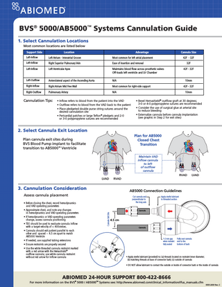
BVS® 5000/AB5000™ Systems Cannulation Guide 1. Select Cannulation Locations Most common locations are listed below Support Side
Location
Cannula Size
Advantage
Left-Inflow
Left Atrium - Interatrial Groove
Most common for left atrial placement
Left-Inflow
Right Superior Pulmonary Vein
Ease of insertion and removal
Left-Inflow
Left Ventricular Apex
Maintains blood flow across prosthetic valves Off-loads left ventricle and LV Chamber
42F - 32F
Left-Outflow
Anterolateral aspect of the Ascending Aorta
N/A
10mm
Right-Inflow
Right Atrium Mid Free Wall
Most common for right-side support
42F - 32F
Right-Outflow
Pulmonary Artery
N/A
10mm
Cannulation Tips:
• Inflow refers to blood from the patient into the VAD • Outflow refers to blood from the VAD back to the patient • Place pledgeted double purse string sutures around the desired cannulation site • Pericardial patches or large Teflon® pledgets and 2-0 or 3-0 polypropylene sutures are recommended
42F - 32F 32F
• Bevel Hemashield® outflow graft at 30 degrees; 3-0 or 4-0 polypropylene sutures are recommended • Consider the use of surgical glue at arterial site to reduce bleeding • Externalize cannula before cannula implantation (see graphic in Step 2 for exit sites)
2. Select Cannula Exit Location Plan for AB5000 Closed Chest Transition
Plan cannula exit sites during BVS Blood Pump implant to facilitate transition to AB5000™ Ventricle
Maintain VAD inflow cannula to left of outflow cannula
RVAD LVAD
3. Cannulation Consideration
AB5000 Connection Guidelines
Assess cannula placement
Cut cannula end perpendicular to the long axis
Apply sterile lubricant to threaded section
Cannula
Cannula Exit Site
• Before closing the chest, record hemodynamics and VAD operating parameters • Approximate chest, and note any changes in hemodynamics and VAD operating parameters • If hemodynamics or VAD operating parameters change, assess cannula positioning • TEE should be used to evaluate cannula inflow with a target velocity of < 400cm/sec. • Cannula should exit patient parallel to each other and spaced ~ 4.5 cm apart to match AB5000 Ventricle • If needed, use supplied tubing extensions • Ensure restraints are properly secured • Use the white threaded cannula restraint marked with a red arrow with the Hemashield ® outflow cannula; use white cannula restraint without red arrow for inflow cannula
LVAD
RVAD
4.5 cm Cannula
(b)
1-2 mm gap when restraint fully seated
Fully seat cannula end to the bottom of barb
(c) (a)
• Apply sterile lubricant (provided) to: (a) threads located on restraint inner diameter; (b) matching threads at base of connector barb; (c) outside of cannula • DO NOT allow lubricant to contact the outside or inside of connector barb or the inside of cannula
ABIOMED 24-HOUR SUPPORT 800-422-8666
For more information on the BVS® 5000 / AB5000TM Systems see: http://www.abiomed.com/clinical_information/ifus_manuals.cfm 0505-2609 Rev. C