Manual
46 Pages
Preview
Page 1
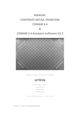
MANUAL CONTRAST-DETAIL PHANTOM CDMAM 3.4 & CDMAM 3.4 Analyser software V2.3
By Marianne Floor and Roeland van der Burght
Artinis Medical Systems Einsteinweg 17 6662 PW Elst The Netherlands Tel: + 31 481 350 980; Fax: + 31 842 105 702
Version 11 - © 2015 Artinis Medical Systems
Manual CDMAM 3.4
Contents ... 2 1
Introduction to the CDMAM ... 4
2
Description of the CDMAM phantom ... 6
3
4
5
6
7
2.1
The aluminium base ... 6
2.2
The PMMA cover ... 7
2.3
The PMMA plates ... 7
2.4
Absorption properties ... 8
2.5
Gold disk specifications ... 8
Directions for use of the CDMAM ... 11 3.1
Making images... 11
3.2
Handling instructions ... 11
3.3
The declaration of conformity ... 11
Evaluation of the CDMAM image ... 13 4.1
Correction scheme... 14
4.2
Correction examples... 14
4.3
Presentation of the results ... 15
CDMAM Analyser ... 17 5.1
Introduction ... 17
5.2
Image readout and scoring ... 17
5.3
Analysis method ... 18
Program structure... 20 6.1
Projects ... 20
6.2
Groups ... 21
6.3
Images... 22
6.4
Analysis ... 23
Analysis results ... 24
-2-
Manual CDMAM 3.4
7.1
Image result ... 24
7.2
Group result ... 25
7.3
Project result ... 26
8
Toolbar ... 29
9
Menu ... 30 9.1
File ... 30
9.2
Edit ... 30
9.3
Project ... 30
9.4
View ... 31
9.5
Window ... 31
9.6
Help ... 31
10
System demands and installation ... 33
10.1
Requirements ... 33
10.2
Use of the USB Key ... 33
11
FAQ ... 34
11.1
Analysis ... 34
11.2
License key ... 34
12
Warranty Policy Artinis Medical Systems B.V. ... 35
13
Literature ... 37
14
Appendices ... 39
-3-
Manual CDMAM 3.4
In mammography it is essential that objects with very small contrast and diameter can be distinguished from the background. Therefore the quality of the technical aspects of mammography equipment should be monitored with regular time-intervals. This is usually performed by measurement of the physical parameters of the X-ray equipment and observation conditions. The main item in quality control however should be the assurance of information transfer from the tissue under examination to the radiologist. Therefore the information content of an image should be monitored.
Figure 1. The CDMAM 3.4 (Contrast-Detail Mammography) phantom.
To evaluate the image quality of mammography systems, a contrast-detail phantom (CDMAM 3.2) was developed (Bijkerk, Lindeijer and Thijssen, 1995). With this phantom, which has been adapted from the Burger-Rose phantom (Burger 1950), the threshold contrast of an imaging system is determined as a function of object diameter, by the detection of pairs of low-contrast objects. In the type 3.2 phantom, the objects (gold disks) ranged in diameter from 0.1 - 3.2 mm and in thickness from 0.05 to 1.6 m, resulting in a The CDMAM-phantom is a result of the project: “Quality Assurance in Mammography, at the Department of Diagnostic Radiology, University Medical Centre Nijmegen, St. Radboud”. It was designed by M.A.O. Thijssen, K.R. Bijkerk and J.M. Lindeijer, in cooperation with the Medical Instrumental Department.
-4-
Manual CDMAM 3.4
radiation contrast range of about 1 - 25 % at standard conditions (molybdenum anode, 30 m molybdenum filtration, 28 kV). The constant improvement of image quality in mammography (high contrast film-screen combinations, digital mammography) made it necessary to visualize lower contrast, as well as a higher spatial resolution (smaller objects) at a higher contrast. The CDMAM phantom was modified to facilitate the evaluation of these systems. In the CDMAM 3.4 version, the gold disks range in diameter from 0.06 to 2.0 mm and in thickness from 0.03 to 2.0 m, resulting in a radiation contrast range of about 0.5 - 30 % at standard mammography exposure conditions (Bijkerk, Thijssen and Arnoldussen, 2000). In this manual, a description of the CDMAM-phantom is given in chapter 1.2, directions for use are presented in chapter 1.3 and the evaluation of the X-ray image is discussed chapter 1.4. In chapter 2 the analyser software is described.
Molybdenum anode, 30 µm Molybdenum filtration, at 28 kV. -5-
Manual CDMAM 3.4
The CDMAM-phantom, shown in figure 1, consists of an aluminium base with gold-disks of various thickness and diameter, which is attached to a PMMA cover. The phantom is delivered with 4 PMMA plates, each with a thickness of 10 mm. The dimensions of phantom and PMMA plates match the standard mammography size (180 x 240 mm).
The aluminium base consists of Al 1050 (99.5% pure aluminium) and has a thickness of 0.5 mm. The base has been polished and was anodized black. Gold-disks (99.99 % pure gold) of variable thickness and diameter have been attached to the base by means of a vaporization technique. The disks are arranged in a matrix of 16 rows and 16 columns (see table 1). The matrix is rotated 45°, to minimize influences of the heel-effect (optical density variations). Within a row the disk-diameter is constant, with an exponential increasing thickness, from 0.03 to 2.00 µm, in steps of approximately 25 and 33 %. The thickness steps were chosen largest in the high contrast part of the phantom, due to the nature of contrast-detail curves of Xray systems (Rose, 1973). Within a column the disk thickness is constant, with an exponential increasing diameter of approximately 25 %, which corresponds with an area step of 50 %.
Column
Thickness [m]
Row
Diameter [mm]
1
0.03
1
0.06
2
0.04
2
0.08
3
0.05
3
0.10
4
0.06
4
0.13
5
0.08
5
0.16
6
0.10
6
0.20
7
0.13
7
0.25
8
0.16
8
0.31
9
0.20
9
0.40
10
0.25
10
0.50
11
0.36
11
0.63
-6-
Manual CDMAM 3.4
12
0.50
12
0.80
13
0.71
13
1.00
14
1.00
14
1.25
15
1.42
15
1.60
16
2.00
16
2.00
Table 1. Thickness and diameter of the gold disks within the phantom Each square contains two identical disks, one in the centre and one in a randomly chosen corner, to allow verification of the detection of each object. Easily memorizable patterns have been avoided. The phantom has been designed such that about half of the disks will be detected by an experienced observer, when state of the art mammography equipment is used at standard exposure conditions.
The cover consists of a 5 mm thick PMMA plate with a cavity with a depth of 2 mm has been milled to accommodate the aluminium base with the gold-disks. The assembly (PMMA + aluminium) has a PMMA-equivalent thickness of 10 mm, under standard mammography-exposure conditions. Between the PMMA cover and the aluminium base a silkscreen printed transparent with X-ray contrasting paint has been placed, showing a grid, the disk thickness and disk diameter. Consequently the X-ray image will show a number of squares arranged in 16 columns and 16 rows, with the disk-diameter shown for each row and the disk-thickness for each column.
The CDMAM-phantom is delivered with 4 PMMA plates, for the simulation of different breast-thicknesses. The plates have a thickness of 10 mm each and are otherwise of the same dimensions as the phantom. For easy identification in the X-ray image of the number and relative position of any added plates, they are marked in one corner with X-ray contrasting paint.
-7-
Manual CDMAM 3.4
Under standard mammography-exposure conditions (Mo-anode, 30 m Mo-filtration, 28 kV), the phantom has a PMMA-equivalent thickness of 10 mm. These conditions will result in an HVL at the image plane of about 0.65 mm Al, when all four PMMA plates are added. This indicates an effective energy of about 19 keV, according to table 2. The linear attenuation coefficient of gold g = 175 mm-1 at this energy (Hubbell, 1982).
Energy [keV]
/ [cm2/g]
HVL [mm Al]
10
25.82
0.099
15
7.836
0.327
20
3.392
0.765
30
1.115
2.300
Table 2. The energy of monochromatic radiation with the mass attenuation coefficient / and the HVL of aluminium. The radiation contrast Cr of the gold disks can be calculated with the formula: 𝐶𝑟 =
𝐼𝑏 −𝐼𝑔 𝐼𝑏
(1)
= 1 − 𝑒 −𝜇𝑔𝑥𝑔
where Ig and Ib represent the X-ray intensities with and without a gold disk and xg the thickness of a gold disk. In table 3 on the next page the radiation contrast of each gold disk is given for standard mammography exposure conditions.
During vaporization of the gold disks, the aluminium plate is covered with a mask with lasered holes of the desired diameter. Due to small irregularities of the mask and specific deviations related to the vaporization process, the disks can deviate slightly from the intended diameter, and will not be exactly circular. Also the thickness of the disks may deviate from the indicated values, mainly due to the gold vaporization profile. Thickness at the centre of the phantom tends to be more, and at the edges less than intended.
-8-
Manual CDMAM 3.4
Column
Thickness [m]
Cr [%]
1
0.03
0.52
2
0.04
0.70
3
0.05
0.87
4
0.06
1.04
5
0.08
1.39
6
0.10
1.73
6
0.13
2.25
7
0.16
2.76
8
0.20
3.44
9
0.25
4.28
10
0.36
6.11
11
0.50
8.38
12
0.71
11.68
13
1.00
16.05
15
1.42
22.00
16
2.00
29.53
Table 3. The radiation contrast Cr of the gold disks, calculated with formula 1, for standard mammography exposure conditions The acceptable magnitude of deviations of gold-disk dimensions is related to the thickness, diameter and area step between two consecutive disk rows and columns in the phantom. This leads to the maximum acceptable deviations, shown in table 4.
Disk dimensions
Minimum step
Acceptable deviation
Diameter
21 %
10 %
Area
42 %
20 %
Thickness
18%
10 %
Table 4.
Maximum acceptable deviation of disk diameter, area and thickness
(proportional to radiation contrast) deviations, related to minimum step size of the disk dimensions.
-9-
Manual CDMAM 3.4
Measurements on a prototype show that most diameter and area deviations are much smaller than the above mentioned specifications. Generally, deviations appear to become more significant at smaller disk diameters (up to 5 % in area and 10 % in diameter). Thickness deviations from indicated values appear to be 10 %.
- 10 -
Manual CDMAM 3.4
To make an X-ray image, the CDMAM-phantom should be positioned on the bucky with the smallest disk-diameters at the thorax side, in combination with one or more PMMA plates. The marks in the PMMA plates should be aligned at the thorax side of the bucky, as is shown in figure 2. Choices have to be made with regard to the exposure technique: Tube potential Focal spot size With or without grid Manual or automatic exposure With or without compression plate For analog mammography: The density of the image has to be checked after the film has been processed. In a series of CD-images, all images should approximately have the same densities in a reference-position on the film.
Store and use the phantom and its belongings at room temperature (15o-25o) and at normal humidity protected for fluid and moisture, dust, etc. preferable in the delivered case. Handle all products with care. PMMA scratches easily which might give that the phantom or plates is useless for evaluation. Clean the materials with non-aggressive general cleaner. The phantom cannot be used in MRI systems or in the neighbourhood of other magnetic materials.
Send back to manufacturer
The Declaration of conformity to the specifications states that the CDMAM 3.4 phantom is in compliance to the norms and product description. By heavy wearing out, misuse or mistreatment of the phantom, the phantom might become unusable or unreliable. In case
- 11 -
Manual CDMAM 3.4
one of the following symptoms is noticed, please contact your local distributor or Artinis Medical Systems. Large scratches and other damage to the body Large scratches and other damage to one of the PMMA Attenuation plates Dents, large scratches or other damage into the aluminium plate The aluminium plate gets loose from the body The grid paint is dissolved by moisture which is visible by high contrast particles at the Xray images and little white speckles between the foil and the body
- 12 -
Manual CDMAM 3.4
CDMAM images can be evaluated at the classical way with human observers or in a digital environment by using the automatic CDMAM Analyser scoring software. The software will be discussed in chapter 5. The (analog) X-ray image of the CDMAM-phantom needs to be evaluated by at least 3 experienced observers. The "Score form CDMAM-phantom" (see Appendix 1) can be used for this purpose. The image should be evaluated in the area where the gold-disks are just visible, by indication of the location of the eccentric disks. At least 3 fields must be observed in each column and each row, in order to comply with the correction scheme, which is described in paragraph 4.1.
Figure 2. Reference configuration of the CDMAM-phantom with the marked PMMA plates on the bucky of a mammography unit
The indicated positions of the eccentric disks have to be compared to the true diskpositions in the phantom, for which the "Evaluation form CDMAM-phantom" can be used (see Appendix 2). To evaluate the observations certain rules have to be applied, taking into account the 4 nearest neighbours (defined by a common vertice) of the field under examination. The evaluation of a particular field must refer to the original observations
- 13 -
Manual CDMAM 3.4
for the nearest neighbours. Examples of the correction scheme are discussed in paragraph 4.2.
In the correction scheme, there are three possibilities for each observation: T: the eccentric disk was indicated at the true position F: the eccentric disk was indicated at a false position N: the eccentric disk was not indicated at all The two main rules within the correction scheme are: A True needs 2 or more correctly indicated nearest neighbours to remain a True. A False or Not indicated disk will be considered as True when it has 3 or 4 correctly indicated nearest neighbours. Exceptions on the two main rules are: A True which has only 2 nearest neighbours (at the edges of the phantom) needs only 1 correctly indicated nearest neighbour to remain True. A False or Not indicated disk which has only 2 nearest neighbours will be regarded True if both nearest neighbours are correctly indicated. The absent corners of the phantom (0.03 m/2.0 mm and 2.00 m/0.06 mm will be regarded True when both nearest neighbours are correctly identified.
Six examples (shown in figure 3) of the correction scheme are discussed below. Example 1:
The common situation. T* remains T because of its 2 correctly indicated
nearest neighbours. F* remains F because it has only 2 correctly indicated nearest neighbours. Example 2:
F* is considered T because it has more than 2 correctly indicated nearest
neighbours. Both T*'s however have only 1 correctly identified nearest neighbour, and thus are considered to be F's. Example 3:
T* remains T because it has 1 out of 2 correctly indicated nearest
neighbours. The absent corner will be considered as an F because it has only 1 correctly indicated nearest neighbour. - 14 -
Manual CDMAM 3.4
T T T T N T* T T N F* T T N N N T
T T T T N T* F* T N N T* T
Figure 3.1
Figure 3.2
F T* T T N N T T N N N T N N N N
F F* T T N T* T T N N T T N N N T
Figure 3.3
Figure 3.4
F F* T* T N N N T N T* N T N N N N
T T* F* T T T T T N T T T N N T T
Figure 3.5
Figure 3.6
N N N T
Figure 3. six examples for the use of the correction scheme.
Example 4:
F* will be considered as a T because of its 2 out of 2 correctly indicated
nearest neighbours. T* will be considered as an F because it has only 1 correctly indicated nearest neighbour. The absent corner will be considered as an F, because it has no correctly indicated nearest neighbours. Example 5:
F* remains an F, because it has only 1 out of 2 correctly indicated nearest
neighbours. Both T*'s are considered as F's because they have none respectively 1 correctly indicated nearest neighbour. Example 6:
T* remains T because it has 1 out of 2 correctly indicated nearest
neighbours. F* will be considered as a T because of 3 correctly indicated nearest neighbours. The absent corner will be considered as a T because of 2 correctly indicated nearest neighbours.
The results can be presented in a logarithmic graph, in which the disk-thickness is plotted against the disk-diameter. The curve through the threshold fields is called the ContrastDetail curve (Thijssen, 1993). The image quality can be expressed in a figure by calculation
- 15 -
Manual CDMAM 3.4
of the ratio of correctly identified disk-positions to the total number of squares (formula 2). 𝑐𝑜𝑟𝑟𝑒𝑐𝑡 𝑜𝑏𝑠𝑒𝑟𝑣𝑎𝑡𝑖𝑜𝑛 𝑟𝑎𝑡𝑖𝑜 =
𝐶𝑜𝑟𝑟𝑒𝑐𝑡 𝑜𝑏𝑠𝑒𝑟𝑣𝑎𝑡𝑖𝑜𝑛𝑠 𝑇𝑜𝑡𝑎𝑙 𝑛𝑢𝑚𝑏𝑒𝑟 𝑜𝑓 𝑠𝑞𝑢𝑎𝑟𝑒𝑠
∙ 100%
(2)
Another method to quantify image quality is called the Image Quality Figure (IQF)-method (Thijssen et al, 1989), which is defined in formula 3. (3)
𝐼𝑄𝐹 = ∑16 1 𝐶𝑖 𝑥 𝐷𝑖,𝑚𝑖𝑛
where Di,min denotes the threshold diameter in contrast-column i. Summation over all contrast-columns yields the IQF. For calculation purposes two extra rules apply: 1. A completely invisible column will result in a D i,min of 2.50 mm (for a gold-disk thickness between 0.03 and 0.25). 2. A completely visible column will result in a Di,min of 0.06 mm (for a gold-disk thickness between 0.16 and 2.0). Image quality increases with an increasing number of correctly identified disk-positions. In this case the IQF will become smaller because the values of diameter and thickness of the threshold-disks are smaller. An alternative method for calculation of the IQFinv has been presented (Thijssen et. al., 2000), in which only the number of partly visible columns of the phantom (n) is taken into account. Furthermore, the summation of is inverted and divided by n, giving a higher IQF at better image quality. The current evaluation method is to determine the threshold thickness per diameter, which results in the following IQF inv calculation: 𝐼𝑄𝐹𝑖𝑛𝑣 = ∑16
100
(4)
𝑖=1 𝑡𝑡ℎ𝑟,𝑖 ∙𝐷𝑖
where tthr,i is the threshold thickness of diameter Di
- 16 -
Manual CDMAM 3.4
The CDMAM Analyser software enables fast automatic readout, scoring and analysis of digital CDMAM 3.4 images. The program uses CDCOM for CDMAM 3.4 for image readout and scoring and analyses the scores according to the described analysis method in the supplement to the European Guidelines fourth edition (Perry et al., 2013). The main change from CDMAM 3.4 Analyser V2.1 to CDMAM 3.4 Analyser V2.2 and higher is the use of threshold thickness converted to human read out and Contrast Detail curves determined by third order polynomial fitting. The analysis results are observer independent. As a prerequisite for reading this manual and using the program correctly the reader should also study the previous chapters on the CDMAM carefully.
For automatic readout and scoring of digital CDMAM images the software tool CDCOM for CDMAM 3.4 is used. The CDCOM software automatically identifies the gold discs on images of the CDMAM phantom and is available from the EUREF website [www.euref.org]. The program attempts to correctly locate the position of the gold discs by first identifying the grid position and reference dots and subsequently the gold discs positions. CDCOM performs the following actions: It reads a single digital CDMAM version 3.4 image and tries to detect the gold disks in the phantom. CDCOM determines in which corner of each cell the gold disc is most likely to be The program checks for each cell if the correct corner was chosen An additional detection step is done using the 3 corners where no marker is present and the centre dot More information regarding CDCOM can be found in the CDCOM Manual. The CDCOM executable can be found in the installation folder of this software (CDCOM.exe). If EUREF releases a new version of CDCOM for CDMAM 3.4 you can download the executable from the EUREF website and replace the current by the new executable file. Please notice you have to give the new executable file the same name.
- 17 -
Manual CDMAM 3.4
The analysis method is according to the Supplement to the European Guidelines. Diameters from 0.1 to 1.0 mm are included during the analysis. The score results of the CDCOM software are averaged and filtered by a Gaussian filter. The probability of gold disc detection by CDCOM is between 25% and 100% by the 4-alternative forced choice method. The gold disc thicknesses having 62.5% detection probability are the threshold thicknesses and are determined by applying psychometric curve fitting by a least squares procedure over the probabilities. The psychometric curve is fitted by (Veldkamp 2003): 𝑝(𝑑) =
0.75 1+𝑒 −𝑓(𝐶−𝐶𝑡 )
(5)
+ 0.25
C = logarithm of signal contrast C = log(1 − e−μd) Ct = signal contrast at the threshold of 62.5% f = fitting parameter p(d) = the probability of detection p(d) of an object with thickness d The attenuation coefficient of gold depends on the beam quality used for the exposure. These values were calculated by DR Dance and KC Young at NCCPM with 3mm PMMA representing the compression paddle, using spectra from Boone (et al., 1997) and attenuation coefficients for materials in the test objects (aluminium, gold, PMMA) from Berger (et al, 2005). In the software these threshold thicknesses are called the Automatic Threshold Gold Thicknesses. The Automatic Threshold Gold Thicknesses are converted to Predicted Human Gold Thicknesses by correction for human read out by 𝑇𝑝𝑟𝑒𝑑𝑖𝑐𝑡𝑒𝑑 = 𝑎𝑇𝑎𝑢𝑡𝑜 𝑛
(6)
Tpredicted =predicted human threshold gold thickness Tauto = Automatic Threshold Gold thickness a, n = fitted parameters with a = 1.441, n = 0.895
- 18 -
Manual CDMAM 3.4
The Fit to Predicted Human Gold Thickness are determined by fitting a third order polynomial through the Predicted Human Gold Thicknesses resulting in the CD (Contrast Detail) Curve by 𝑏
𝑐
𝑑
𝑥
𝑥
𝑥
(7)
𝑇𝑓𝑖𝑡 = 𝑎 + + 2 + 3 Tfit = Fit to Predicted Human Gold Thickness (um) x = detail diameter (mm) a, b, c and d = coefficients adjusted to achieve a least squares fit, and are ≥ 0 The IQFinv number is computed according to 𝐼𝑄𝐹𝑖𝑛𝑣 = ∑𝑛
100
(8)
𝑖=1 𝑡𝑖,𝑡ℎ𝑟 ∙𝐷𝑖
where ti,thr denotes the threshold thickness (Fit to Predicted Human Gold Thickness) in diameter-column i. The thickness is given in µm whereas the diameter is taken in mm. The total detected (%) is determined by 𝑇𝑜𝑡𝑎𝑙 𝑑𝑒𝑡𝑒𝑐𝑡𝑒𝑑 (%) =
𝑛𝑢𝑚𝑏𝑒𝑟 𝑜𝑓 𝑑𝑒𝑡𝑒𝑐𝑡𝑒𝑑 𝑔𝑜𝑙𝑑 𝑑𝑖𝑠𝑐𝑠 𝑡𝑜𝑡𝑎𝑙 𝑛𝑢𝑚𝑏𝑒𝑟 𝑜𝑓 𝑔𝑜𝑙𝑑 𝑑𝑖𝑠𝑐𝑠
× 100%
- 19 -
(9)
Manual CDMAM 3.4
The data and images in the program are organized in projects with groups (Figure 4). The project may contain several groups, each having by example other settings during imaging of the phantom, allowing easy comparison of the influence of the settings to the image quality. A group should contain multiple images made with the same settings. We recommend to use at least 8 images, but preferable 16 or more per group for reliability of the results. The readout and score result can be viewed per project, group or image. Analysed images are marked with a green sign. In case the analysis failed, the image is marked with a yellow question mark, mostly caused by wrong or incomplete DICOM header information (see chapter 6.3).
Figure 4. Program structure
A new project can be made via the menu, File, New project. Existing projects can be opened via menu, File, Open project or via the toolbar opening button. The project can be saved via menu, File, Save project (as) or via the toolbar saving button, which also changes the project name from the standard “new project” to the project file name. Latest projects can be opened via menu, File. The project has properties, which can be viewed via menu, Project, properties or via the right mouse button at the project. The first tab offers to add comments for project description and the second to use relative file paths or changing the setting if DICOM files are opened or not by addition to a project. The relative file paths save the image file paths relatively to the project file path, which is easy for copying project with images from a computer to another computer, USB or CD by example.
- 20 -